Brunetorhynchus deconincki

Description:
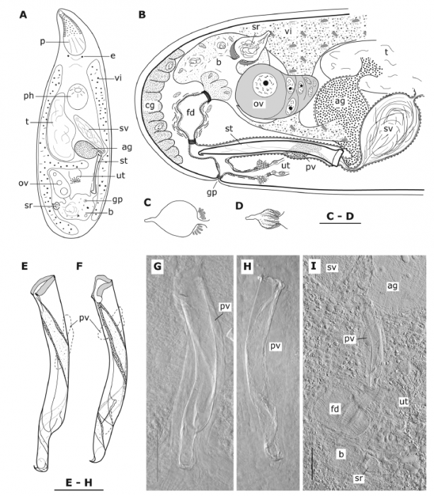
Brunetorhynchus deconincki n. gen. n. sp. (A) The living animal as drawn by M. Brunet. (B) Reconstruction of the atrial organs from sagittal sections, seen from the right. (C, D) The seminal receptacle in two individuals (from whole mounts). (E–H) The stylet: (E, G) of the holotype, (F) of another individual from the Bay of Marseille, (H) of an individual from Calvi. (I) Some atrial organs as seen in the whole mount of a third individual from the Bay of Marseille. Note the globular aspect of the female duct in this specimen and the longitudinal muscles in bundles. (scale bars = 25 µm)
Included On The Following Pages:
- Life
- Cellular
- Eukaryota (eukaryotes)
- Opisthokonta (opisthokonts)
- Metazoa (animals)
- Bilateria
- Protostomia (protostomes)
- Spiralia (spiralians)
- Platyhelminthes (flatworms)
- Rhabdocoela
- Polycystididae
- Brunetorhynchus
- Brunetorhynchus deconincki
- Rhabditophora
This image is not featured in any collections.
Source Information
- license
- cc-by-nc-sa-4.0
- copyright
- WoRMS Editorial Board
- contributor
- Artois, Tom [email]
- original
- original media file
- visit source
- partner site
- World Register of Marine Species
- ID