Image of Maxillopoda

Description:
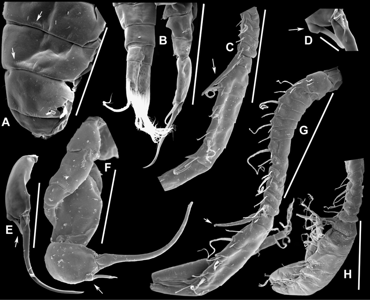
Figure 40.Notodiaptomus coniferoides male, SEM photographs. A Pedigers 2–5 (200 µm) B Last 3 urosome somites, CR, and P5R (300 µm) C Segments 9–18 of A1R (200 µm) D Detail showing insertion of lateral spine on Exp2P5R (20 µm) E Exp2P5R (100 µm) F P5 (100 µm) G A1R (300 µm) H A1R from segment 7 to tip (200 µm).
Included On The Following Pages:
- Metazoa (animals)
- Arthropoda (arthropods)
- Maxillopoda
- Notodiaptomus
- Calanoida (Calanoid copepods)
- Diaptomidae
- Notodiaptomus coniferoides
This image is not featured in any collections.
Source Information
- license
- cc-by-3.0
- copyright
- Gilmar Perbiche-Neves, Geoffrey Allan Boxshall, Daniel Previattelli, Marcos Gomes Nogueira, Carlos Eduardo Falavigna da Rocha
- bibliographic citation
- Perbiche-Neves G, Boxshall G, Previattelli D, Nogueira M, da Rocha C (2015) Identification guide to some Diaptomid species (Crustacea, Copepoda, Calanoida, Diaptomidae) of “de la Plata” River Basin (South America) ZooKeys (497): 1–111
- original
- original media file
- visit source
- partner site
- Zookeys
- ID