Image of Argyrodiaptomus falcifer (Daday 1905)

Description:
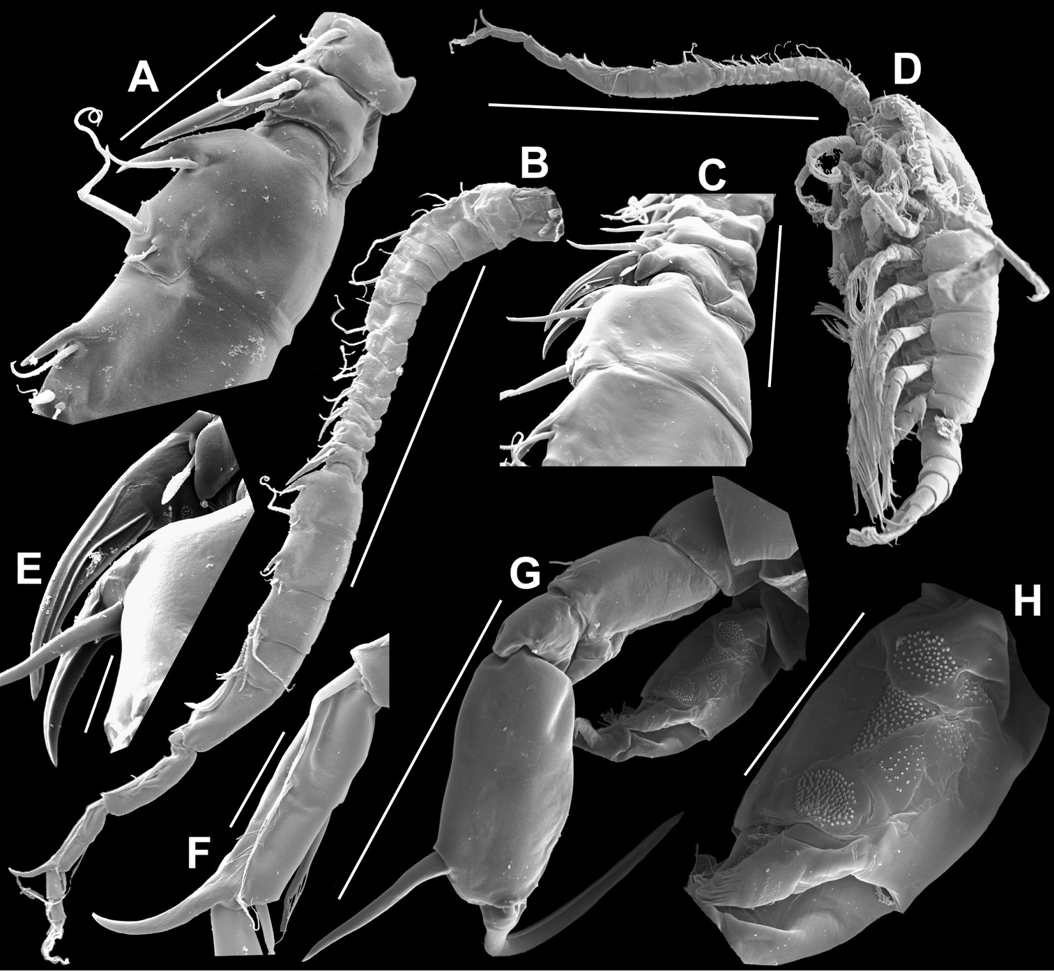
Figure 16.Argyrodiaptomus falcifer male, SEM photographs. A Segments 12–15 of A1R, showing detail of spinous process on segment 14 (arrowed) (100 µm) B Entire A1R (500 µm) C Segments 11–15 of A1R (100 µm) D Adult male, lateral view (1000 µm) E Spinous processes on segments 13 and 14 A1R (20 µm) F Segment 20 of A1R, showing falciform process (50 µm) G Complete P5, caudal view (200 µm) H Medial surface of BspP5L, showing spinular ornamentation (50 µm).
Included On The Following Pages:
- Metazoa (animals)
- Diaptomidae
- Argyrodiaptomus falcifer
- Life
- Cellular
- Eukaryota (eukaryotes)
- Opisthokonta (opisthokonts)
- Bilateria
- Protostomia (protostomes)
- Ecdysozoa (ecdysozoans)
- Arthropoda (arthropods)
- Pancrustacea
- Multicrustacea (typical crustaceans)
- Copepoda (copepods)
- Calanoida (Calanoid copepods)
- Argyrodiaptomus
- Hexanauplia
- Panarthropoda
This image is not featured in any collections.
Source Information
- license
- cc-by-3.0
- copyright
- Gilmar Perbiche-Neves, Geoffrey Allan Boxshall, Daniel Previattelli, Marcos Gomes Nogueira, Carlos Eduardo Falavigna da Rocha
- bibliographic citation
- Perbiche-Neves G, Boxshall G, Previattelli D, Nogueira M, da Rocha C (2015) Identification guide to some Diaptomid species (Crustacea, Copepoda, Calanoida, Diaptomidae) of “de la Plata” River Basin (South America) ZooKeys (497): 1–111
- original
- original media file
- visit source
- partner site
- Zookeys
- ID