Image of Bryobia cinereae Auger & Migeon 2014

Description:
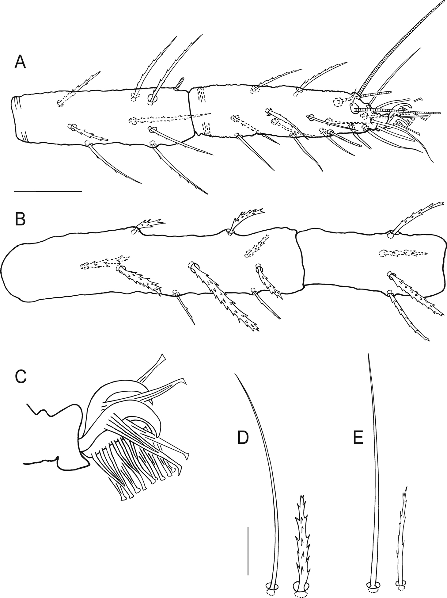
Figure 3.Bryobia belliloci sp. n., female: A tarsus and tibia I B genu and femur I C claws and empodia I–IV D coxisternal setae 1b and 1c E Bryobia cinereae Auger & Migeon (2014), Holotype female, coxisternal setae 1b and 1c. Scale bars = 25 µm (A–B), 10 µm (C–E).
Included On The Following Pages:
- Metazoa (animals)
- Arthropoda (arthropods)
- Arachnida (arachnids)
- Prostigmata (prostigmatan mites)
- Tetranychidae (spider mites)
- Bryobia
- Bryobia cinereae
- Life
- Cellular
- Eukaryota (eukaryotes)
- Opisthokonta (opisthokonts)
- Bilateria
- Protostomia (protostomes)
- Ecdysozoa (ecdysozoans)
- Chelicerata (chelicerates)
- Acari (mites)
- Acariformes (mites and ticks)
- Trombidiformes
- Eleutherengona
- Raphignathina
- Tetranychoidea
- Panarthropoda
This image is not featured in any collections.
Source Information
- license
- cc-by-3.0
- copyright
- Philippe Auger, Tea Arabuli, Alain Migeon
- bibliographic citation
- Auger P, Arabuli T, Migeon A (2015) Two new species of Bryobia (Acarina, Prostigmata, Tetranychidae) from South France ZooKeys (480): 21–39
- original
- original media file
- visit source
- partner site
- Zookeys
- ID