Tridax Dicot stem

Description:
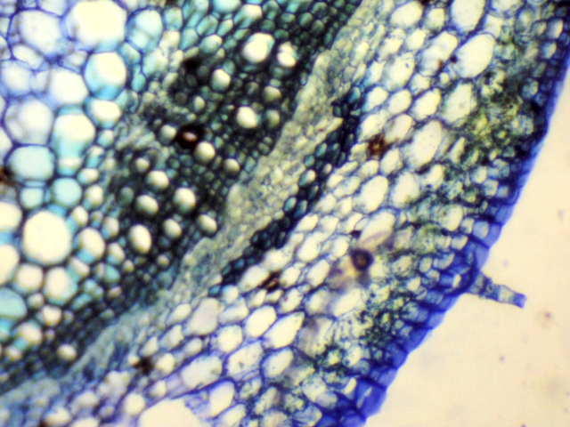
Summary[edit] Description: English: This methylene blue-stained Tridax stem section shows the 4 types of plant tissues. The bigger cells appear at the top left of the image was Ground tissue. The thick-walled cells at the center of the image were Vascular tissue. The outer most layer of the stem appears at the bottom left of the image. Lateral Meristematic tissue with thin-walled small size cells present in a row. Аҧсшәа: మిథిలిన్ బ్లూ తో రంజనం చేయబడిన ఈ గడ్డి చామంతి మొక్క కాండం అడ్డుకోత చేదనం మొక్కలలలోని నాలుగు రకాల కణజాలాలను చూపుతుంది.చిత్రంలో పైన ఎడమ వైపున ఉన్న పెద్ద కణాలు సంధాయక కణజాలం.చిత్రంలో మద్యలో కనిపిస్తున్న మందమైన కవచాలు గల కణాలు ప్రసరణ కణజాలం.అన్నింటికంటే వెలుపల కాండాన్ని చుట్టి ఉన్కణాల వరుసలు త్వచ కణజాలం. విభజ్య కణజాలం చిన్న చిన్న కణాలతో పలుచని కవచాలతో ఒక వరుసలో ప్రసరణ కణజాలం మధ్యనుండి ఉంది. Date: 23 July 2019, 11:43:02. Source: Own work. Author: Krishna satya 333. This work is for energizing the Andhra Pradesh SCERT biology textbook 9th class, 2 nd lesson, activity 7. Licensing[edit] : This file is licensed under the Creative Commons Attribution-Share Alike 4.0 International license. :. You are free: to share – to copy, distribute and transmit the work to remix – to adapt the work Under the following conditions: attribution – You must give appropriate credit, provide a link to the license, and indicate if changes were made. You may do so in any reasonable manner, but not in any way that suggests the licensor endorses you or your use. share alike – If you remix, transform, or build upon the material, you must distribute your contributions under the same or compatible license as the original. https://creativecommons.org/licenses/by-sa/4.0 CC BY-SA 4.0 Creative Commons Attribution-Share Alike 4.0 truetrue.
Included On The Following Pages:
- Life
- Cellular
- Eukaryota (eukaryotes)
- Archaeplastida (plants)
- Chloroplastida
- Streptophyta
- Embryophytes
- Tracheophyta (vascular plants)
- Spermatophytes
- Angiosperms
- Eudicots
- Superasterids
- Asterids
- Asterales
- Asteraceae (composite family)
- Tridax (tridax)
This image is not featured in any collections.
Source Information
- license
- cc-by-sa-3.0
- copyright
- Krishna satya 333
- creator
- Krishna satya 333
- original
- original media file
- visit source
- partner site
- Wikimedia Commons
- ID