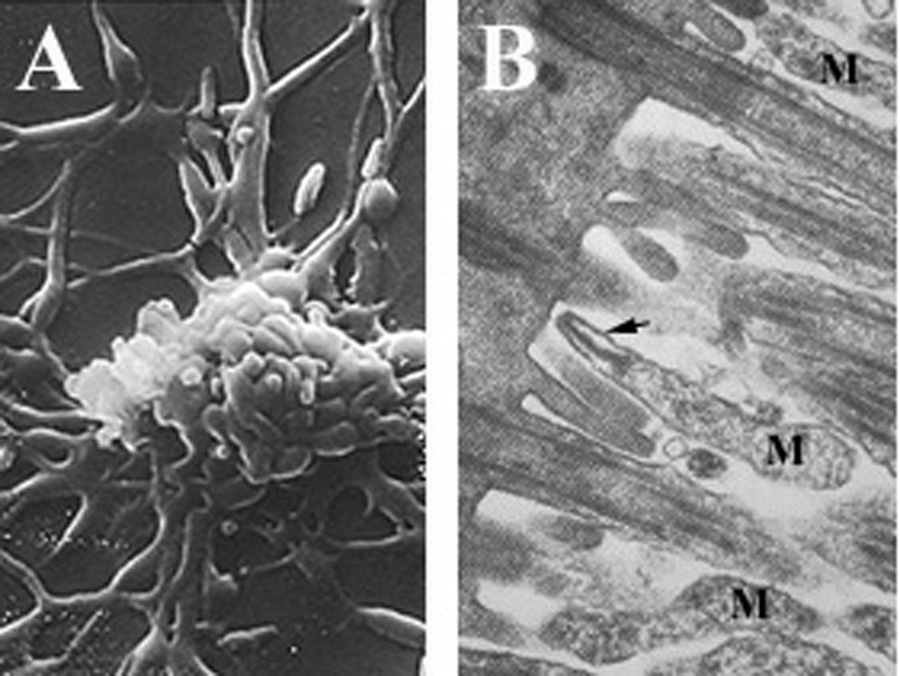
Mycoplasma pneumoniae cells attached to ciliated mucosal cells

Description:
Summary[edit] Description: English: "A, scanning electron microscopy of filamentous M. pneumoniae. B, transmission electron microscopy of flask-shaped M. pneumoniae (M) attached by the terminal tip organelle (arrow) to ciliated mucosal cells. Magnification: A, x10,000;B, x36,000.". Date: 22 April 2002, 12:50:45. Source: Rottem, S., S., N., and D., J. (2012) in Biomedical Tissue Culture (Ceccherini-Nelli, L., ed.), InTech [online] http://www.intechopen.com/books/biomedical-tissue-culture/contamination-of-tissue-cultures-by-mycoplasmas (Accessed December 3, 2013). Author: Rottem et al.
Included On The Following Pages:
- Life
- Cellular
- Bacteria
- NO NAME!
- Tenericutes
- Mollicutes
- Mycoplasmatales
- Mycoplasmataceae
- Mycoplasma
- Mycoplasma pneumoniae
This image is not featured in any collections.
Source Information
- license
- cc-by-3.0
- copyright
- Rottem et al.
- original
- original media file
- visit source
- partner site
- Wikimedia Commons
- ID