Isoaulactinia stelloides

Description:
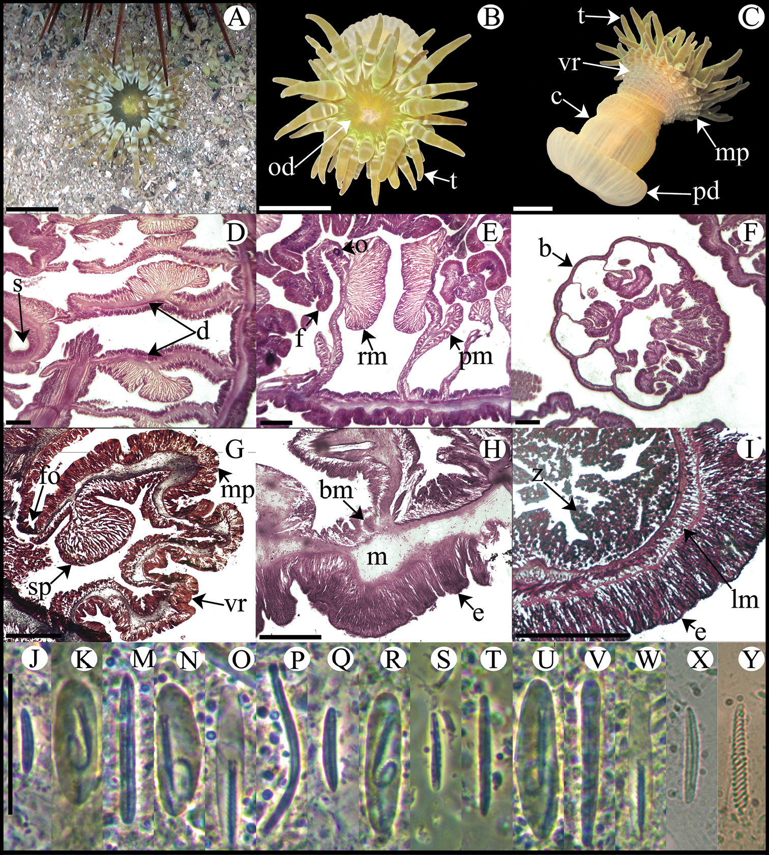
Isoaulactinia stelloides. A Live specimen in natural habitat B Oral view C Lateral view D Detail of directives showing a siphonoglyph E Cross section through proximal column F Detail of brooded juvenile G Longitudinal section through margin showing marginal sphincter muscle and marginal projection H Longitudinal section though base showing basilar muscles I Cross section through tentacle J–Y Cnidae.– marginal projection: J small basitrich K macrobasic p-mastigophore; actinopharynx: M basitrich N macrobasic p-mastigophore O microbasic p-mastigophore P long, curved basitrich; column: Q small basitrich R macrobasic p-mastigophore; filament: S small basitrich T basitrich U macrobasic p-mastigophore V microbasic b-mastigophore W microbasic p-mastigophore; tentacle: X basitrich Y spirocyst. Abbreviations.– b: brooded juvenile, bm: basilar muscle, c: column, d: directives, e: epidermis, fo: fosse, lm: longitudinal muscle, m: mesoglea, mp: marginal projection, o: oocyst, od: oral disc, pd: pedal disc, pm: parietobasilar muscle, rm: retractor muscle, s: siphonoglyph, sp: sphincter, t: tentacle, vr: verrucae, z: zooxanthellae. Scale bars: A–C: 10 mm; D–I: 200 μm; J–Y: 25 μm.
Included On The Following Pages:
- Life
- Cellular
- Eukaryota (eukaryotes)
- Opisthokonta (opisthokonts)
- Metazoa (animals)
- Cnidaria (cnidarians)
- Anthozoa (anemones and corals)
- Actiniaria (sea anemones)
- Actinioidea
- Actiniidae
This image is not featured in any collections.
Source Information
- license
- cc-by-3.0
- copyright
- González-Muñoz R, Simões N, Tello-Musi J, Rodríguez E
- creator
- González-Muñoz R, Simões N, Tello-Musi J, Rodríguez E
- original
- original media file
- visit source
- partner site
- Wikimedia Commons
- ID